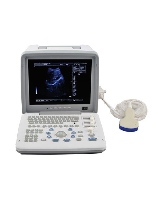
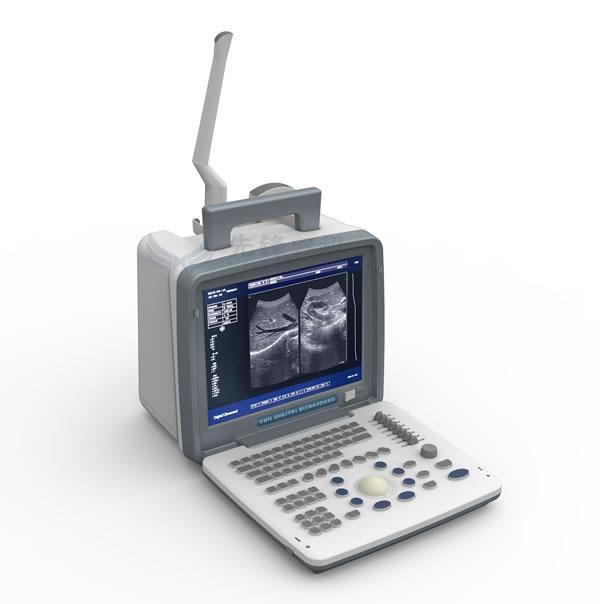
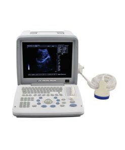
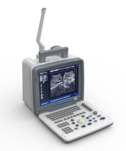

Portable LED screen ultrasound
XF300 (LED) B ultrasound diagnose medical instrument
The fully digital ultrasound image diagnostic equipment,
- Standard configuration: 12in LCD screen main unit with one probe
- user?s manual
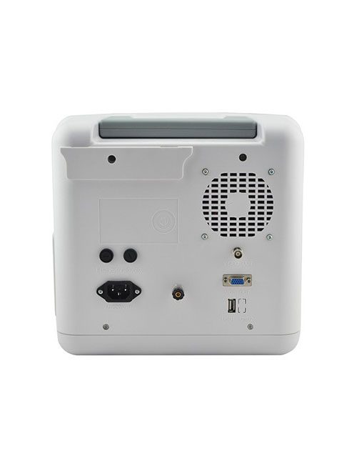
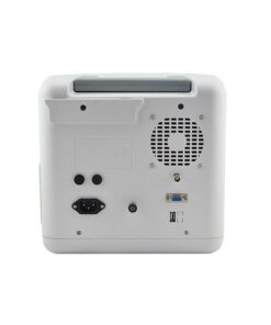
- USB,VGA,VIDO Print port
- two probe sockets
- 18 months warranty
- 110-240 voltage
- Automatic patient?s report
- M, B/M, BB, 4B, B mode
- Optional: convex array probe
- trans-vaginal probe
- high fequency linear probe
- micro-convex array probe
- ?
- Main features: Excellent value for money Full digital imaging technology DBF: Digital Beam Forming RDA: Real-time dynamic aperture imaging DRA: Dynamic real-time acoustic apodizer DRF: Dynamic receiving focus DFS: Dynamic frequency scanning Ensure that the image is not distorted, edge clearer, level richer Humanized operating design 8 sections TGC gain adjustment With abundant built-in software packages Comprehensive software measurement capabilities, to allow more extensive clinical application Two probe/three probeconnectors (optional) Probe connected with a variety of choice to achieve maximum functionality VLSI, advanced technology, stable performance Reasonable cost control, to achieve the perfect embodiment of price and performance
- Specifications: Image mode: B, B/B, 4B, B+M, M Image magnification: *0.8, *1.0, *1.2, *1.5, *1.8, *2.0 Local zoom: 2 times local zoom in real time Dynamic range: 64dB ? 96dB adjustable Focus: 4-segments dynamic electronic focuses selected Gray scale: 256 Pre-processing: variable aperture, image direction, dynamic filter, edge enhancement, etc. Post-processing: digital space time filter, 8 y corrections, 16 Pseudo Colors, column correlation, frame correlation, spot correlation, linear interpolation, etc. Multi-frequency: 2.5MHz/3.0MHz/3.5MHz/4.0MHz etc. Multi-frequency selected Measurement: distance, circumference/acreage, HR, pregnancy week (BPD, GS,CRL,FL, AC) and calculating fetus weight, etc. Annotation: Chinese/English interface transition; hospital name, doctors/patients name, case number, gender, age; 16 body marks with probe location, full screen character annotation, real time clock display Puncture lead: Image B: Puncture guide line under B mode Gain adjustment: 8-segments TGC adjustment, GAIN adjustment or near field, far field, overall gain adjustable Image reverse: left/right, black/white, up/down Storage: 128 images permanent storage Cine loop: 256 images real time display cycling/one-by-one checked Output interface: 2 SVGA video outputs, SVGA color monitor circumscribed; 2 PAL video outputs, which can be connected with PAL standard monitor, Video thermal recorder, ultrasound image workstation, etc. Optional features: multiple probes connectable, USB port, Video thermal recorder Probe connectors: 2 socket, multiple probes connectable
- ?: ?
- Standard: Main unit 60R/3.5MHZ Convex array probe USB 2.0, VGA port Video connector
Real photos
Factory line
Related products

SPECIALS
R96,605.00 Ex VAT